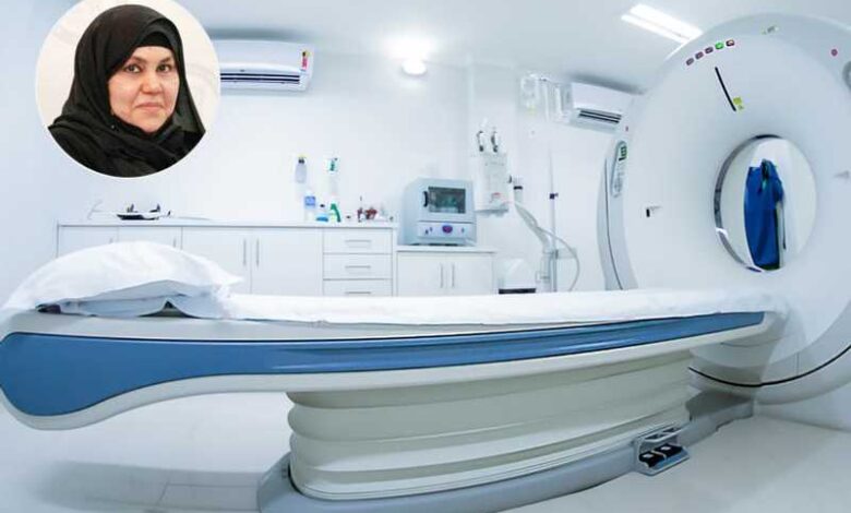
Nuclear medicine imaging is an examination that provides images or scans of internal body parts using small amounts of radioactive material. This examination is used to show images of the body’s organs and parts, which cannot be seen well using usual x-rays, and it particularly allows viewing of many cases of abnormal tissue growth, such as tumors. A radiologist who specializes in using X-rays and other imaging techniques details the examination.
Despite their importance, many people hesitate to undergo these tests, and the reason may be the lack of knowledge about what is done during these tests or the name’s association with nuclear, reports Al-Qabas daily.
According to Dr. Fatima Jassab Al-Saidi, a professor in the Department of Nuclear Medicine – Faculty of Medicine at Kuwait University, nuclear medicine is a branch of medicine that uses the nuclear properties of radioactive materials with the aim of studying organ functions, metabolism, and diagnosing diseases. It also includes therapeutic and research applications.
She went on to say, “The basis of nuclear medicine is based on the use of radioactive pharmaceutical substances (which have nuclear atoms) with a short half-life (meaning that the rays they emit have a short lifespan), and they are labeled with a substance present in the body or needed by the body, as it is metabolized by the organ or area to be examined or treat it.”
Here the doctor answers the 5 most common questions about nuclear medicine examinations:
- How do radioactive materials enter the body?
Radioactive material can be introduced into the body through several methods, such as: inhalation — intravenous injection, which is the most common. – Subcutaneous injection – Intra-abdominal injection — ingestion.
After the radioactive drug enters the body, the radioactive materials will be concentrated in the area or tissue to be examined, and then rays will be emitted from inside the patient, and these radioactive emissions can be measured and photographed using a special camera or imaging device that generates images and provides accurate information.
2. What are the diagnostic methods for nuclear medicine examinations?
These methods vary, the most common are: Scintigraphy using an Anger or gamma camera; Positron Tomography (PET); Medical imaging with single flash gamma ray CT (SPECT); Scintigraphy combined with other imaging techniques, such as MRI or CT.
3. What happens during a nuclear medicine imaging procedure?
A small dose of the radioactive material is given in the form of an injection or a solution that is swallowed, and this depends on the area of the body to be examined and the type of scan.
Also, depending on the type of scan, imaging may be performed immediately or the patient may be asked to return for the actual scan several hours later, or a day after ingesting the radioactive material.
The patient is asked to lie on a special table during the scanning, where the camera used in nuclear medicine imaging is fixed over the area to be examined, while the patient remains relaxed and calm and does not feel anything during the examination, which lasts from 15 minutes to approximately an hour, after which he can leave for the home.
4. How will I feel during and after the medical procedure?
The possibility of radiopharmaceutical injections causing allergic reactions or negative symptoms is very low.
When injected into a vein, you will feel a slight tingling sensation when the needle is inserted into the vein, and you may also feel cold when the solution is injected.
If the method of introducing radioactive materials is swallowing, they may have a slight taste. As for inhalation, you should not feel any different than when you inhale the air around you or hold your breath.
Nuclear imaging itself does not cause pain, and is very safe.
5. When and how does radioactive material exit the body?
Over time, radioactive materials lose their radioactivity. These substances can also be excreted from the body through sweat and urine during the first hours or days following the test.
Therefore, the patient is advised to drink a lot of water to speed up the exit of the radioactive material from the body.
Giving important information, Dr Al-Saidi said nuclear medicine imaging allows many cases of abnormal tissue growth, such as tumors, to be seen. It is an examination that provides images of the internal parts of the body using small amounts of radioactive materials.
Radioactive materials come out during the first hours after the examination through sweating, urine, or excretion.
Radioactive material can be introduced into the body through several methods, such as: inhalation and intravenous injection.


